
MDC Researchers Discover Molecule Responsible for Axonal Branching
Through the ramification of its fiber-like axon, a single neuron can send branches and thus transmit information into several target areas at the same time. In principle, neurobiologists distinguish between two kinds of axonal branching: branching of the growth cone at the tip of an axon and the sprouting of collaterals (interstitial branching) from the axon shaft.
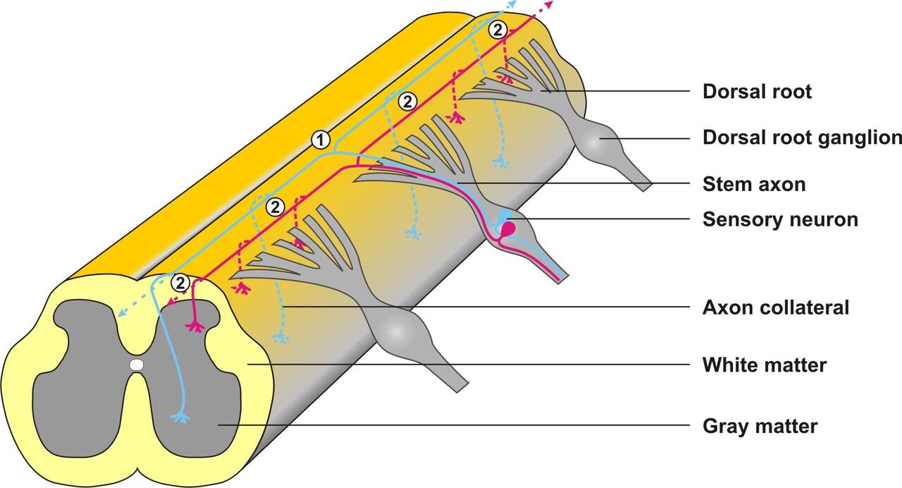
Schematic representation of a sensory neuron. When the axon of the sensory neuron grows into the gray matter of the spinal cord, two types of branching can be observed: At the dorsal root entry zone the axon shaft divides into two branches (1), which continue to grow on the surface of the spinal cord in opposite directions. Out of these branches collaterals then sprout in several places (2) thus enabling the transmission of a signal to several target cells. (Drawing: Hannes Schmidt / Copyright: MDC)

The blue color shows the dispersion area of CNP in a mouse embryo twelve and a half days after fertilization. Through alterations via genetic engineering the original gene for CNP was replaced by the lacZ gene in the depicted mouse. In a color reaction the expression of CNP in the tissue can thus be made visible. (Photo: Hannes Schmidt / Copyright: MDC)
Both forms of axonal branching can be observed in sensory neurons, which transmit the sensation of touch, pain and temperature, among others. When the axons of these neurons reach the spinal cord, their growth cones first split (bifurcate) and consequently the axons divide into two branches growing in opposite directions. Later new
branches sprout from the shaft of these daughter axons which penetrate the gray matter of the spinal cord.
Through investigations on sensory neurons, Dr. Hannes Schmidt and his colleagues were able to identify a protein which triggers the splitting of the growth cone of
the sensory axons: the peptide CNP (the abbreviation stands for C-type natriuretic peptide). In transgenic mice the scientists were able to show that CNP is formed in the
spinal cord precisely when sensory neurons grow into it. In the absence of CNP bifurcation can no longer occur which results in reduced neuronal connectivity in the spinal cord.
The new findings supplement earlier discoveries of the research group of Professor Rathjen according to which a cGMP-signaling cascade is responsible for the bifurcation of sensory axons. When CNP binds to its receptor Npr2 (natriuretic peptide receptor 2) on the surface of the axons, this signaling cascade is set in motion, which in turn induces the formation of the secondary messenger molecule cGMP. This messenger molecule then activates the protein kinase cGKI (cGMP-dependent protein kinase I), which can switch on and off a whole series of target proteins. The cytoskeleton of the neurons is thus altered in such a way that their growth cone splits into two daughter axons.

Dorsal view of the spinal cord with single visible sensory neurons A) Wild-type with bifurcations marked by arrows and B) CNP knock-out mouse. (Photo: Hannes Schmidt / Copyright: MDC)
Next, the researchers want to identify these target proteins. Further analyses should clarify whether the cGMP signaling cascade likewise regulates the branching of other
axon systems and whether this impacts the sensation of pain.
*C-type natriuretic peptide (CNP) is a bifurcation factor for sensory neurons
Author affiliation: Hannes Schmidta, Agne Stonkutea, René Jüttnera, Doris Koeslingb, Andreas Friebeb,c, Fritz G. Rathjena
a Department of Developmental Neurobiology, Max Delbrück Center for Molecular Medicine, Robert Rössle Str. 10, D-13092 Berlin
b Institute for Pharmacology and Toxicology, Ruhr University Bochum, D-44780 Bochum
c Present address: Institute for Physiology I, University of Würzburg, Röntgenring 9, D-97070 Würzburg
Correspondence to F.G. Rathjen: rathjen@mdc-berlin.de
Barbara Bachtler
Press and PublicAffairs
Max Delbrück Center for Molecular Medicine (MDC) Berlin-Buch
Robert-Rössle-Straße 10
13125 Berlin, Germany
Phone: +49 (0) 30 94 06 - 38 96
Fax: +49 (0) 30 94 06 - 38 33
e-mail: presse@mdc-berlin.de
http://www.mdc-berlin.de/


