
µComputed Tomography (µCT)
Computed Tomography (CT) is an extremely useful imaging technique, suitable for diverse biomedical applications. It is commonly used for diagnostic purposes in clinical practice and has been also successfully integrated in preclinical research. Here, due to the small size of the object under investigation (sample or animal), not only is the non-destructive imaging of the whole volume possible, but spatial resolution at the micro-meter level can be also easily achieved. Hence, we speak of microCT (µCT).
Our scanners

- Imaging Principle
An object is imaged with X-rays from multiple angles to acquire projection images and subsequently use them to reconstruct a 3D representation (3D model) of that object. The contrast in obtained images results from different absorption levels of the X-rays by different tissue types. Therefore, dense elements absorbing high amounts of X-rays are well visible in the image while surrounded by less absorbing background. Similarly, an organ absorbing only very little X-rays (e.g. lungs) will be well contrasted by the surrounding soft tissues.
- Image Acquisition and Analysis
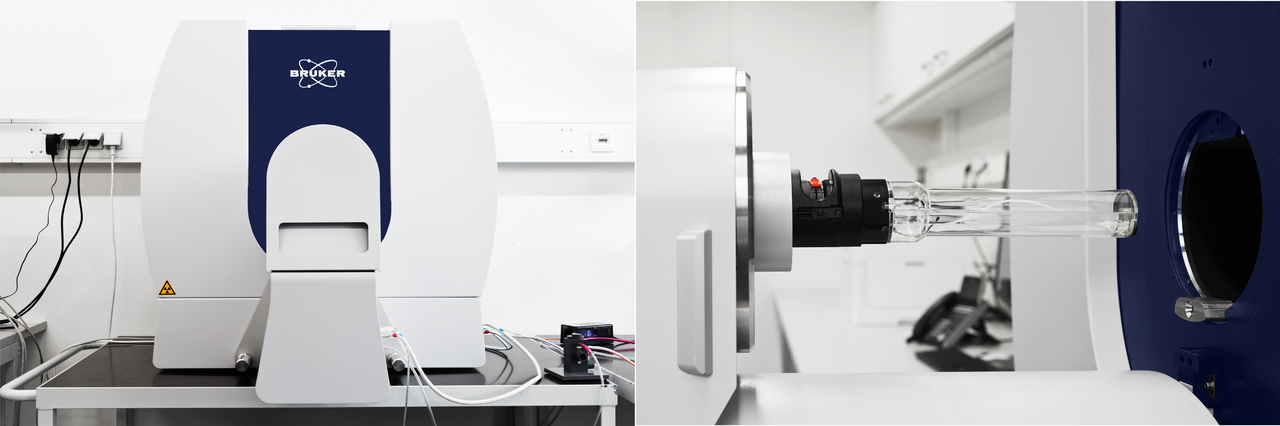
In SkyScan 1276, the object under investigation is positioned on a sample holder of a size adequate to the sample dimensions, or in an animal cassette for in vivo scanning. The X-ray source and X-ray detector rotate on the gantry around the object.
In SkyScan 1272, the sample is positioned on a stage which rotates very slowly around its axis during image acquisition. The X-ray source and the X-ray detector are fixed in the machine.
Both scanners are equipped in different X-ray filters and image magnification can be adjusted as required. This flexibility of the devices allows us to optimize the image acquisition parameters for different objects. Consequently, small samples can be imaged with a very high spatial resolution, allowing visualization of elements as tiny as bone trabeculae, developing organs in embryos, or glomeruli in a mouse kidney. Whereas in vivo studies can be performed using a minimal X-ray dose.
Furthermore, we pay close attention to using the most suitable tool for image analysis. 3D rendering is usually performed with CTVox or Amira, and the quantitative image analysis with CTAn, Amira, or ImageJ. We are also open for further developments.
- µCT Applications
The main applications of µCT are bone and lung imaging. However, if a contrast agent is used, investigations of other body organs and soft tissues are also well possible. Imaging can be conducted either in vivo (with X-ray dose monitoring) or ex vivo, depending on the study question. During in vivo experiments, monitoring of physiological parameters, such as body temperature and breathing rate, is performed alongside the image acquisition. Due to this flexibility, a broad range of research fields can benefit from µCT, including oncology, pulmonology, cardio-vascular research, developmental biology, neurology and other.
- Examples
The primary application of µCT in biomedical research is bone imaging. Due to its high density, bone tissue provides strong signal and excellent contrast in obtained images. This is the basis for the subsequent image analysis. Thus, not only 3D visualization, but also a quantitative analysis (e.g. bone morphometry or bone mineral density measurement) is easily possible.
Bones of a rat front paw scanned ex vivo. (Sample provided by the reasech group of Enno Klußmann, MDC).
Whole body imaging of a mouse or a rat can be performed with or without a contrast agent, depending on the main focus of the study. Thus, dimensions, proportions and orientation of body organs can be then studied in their natural positions. Despite the large filed of view, spatial resolution remains at the µm level.
During in vivo scaning, the animal is anaesthetized and the radiation dose, as well as physiological parameters, are monitored.
Whole body mouse image, acquired in vivo with a contrast agent.
Vascular imaging of different body organs is performed after a perfusion with a hardening contrast agent. Blood vessels with the diameter as small as 20 µm can be reliably imaged and analzed.
Coronary blood vessels of a rat heart, imaged after perfusion with a hardening contrast agent. (Sample provided by the research group of Ralf Dechend & Dominik Müller, MDC).
Since lungs contain a lot of air, they have a low physical density and absorb only a small fraction of the X-rays. They appear dark in a CT image, contrasting the surrounding tissues. This high contrast allows volumetric measurements of the lungs at different stages of the breathing cycle and hence, supports not only structural, but also funtional studies in the field of pulmunology and oncology.
Mouse lungs imaged in vivo throughout a breathing cycle.